Canine Leukemia: A Case Study
May 19, 2021Written by: Katie Cummings, Veterinary Technician Student
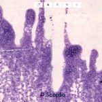
A 10-year-old female spayed boxer mix presented to The Oncology Service for evaluation of lymphocytosis and peripheral lymphadenopathy following acute, significant vomiting, diarrhea and inappetence treated by the referring veterinarian. Cytology of peripheral lymph nodes, as well as a complete blood count and blood chemistry panel were sent to the referrer’s laboratory, diagnosing lymphoma on cytology and lymphocytosis on pathologist’s review of the CBC. She also had a history of copper storage disease.
Examination
On examination by the oncologist, she was bright, alert, and responsive, the vomiting and diarrhea had ceased, but peripheral lymph nodes were markedly enlarged, some as large as 4 centimeters in length. The remainder of her physical exam was unremarkable. Owners elected to move forward with flow cytometry of the peripheral blood, with the plan of treating that disease and hoping her lymph nodes respond as well. With flow cytometry pending, Prednisone was initiated at the dose of 40mg/m2 q24 for 14d, then every other day until otherwise recommended.
Flow cytometry interpretation of the peripheral blood by Dr. Anne Avery, Professor of Immunology at Colorado State University: “The flow cytometry study revealed a homogeneous population of CD4 T cells. This finding is diagnostic for T cell lymphoma/leukemia. We do not have prognostic information for this particular phenotype when diagnosed in the peripheral blood.”


Recommendations
Due to the lack of information in prognosis and treatment information for her specific type of T-cell leukemia, owners elected to pursue oral lomustine treatment with the hope that if she responds well a 4–6-month period of great quality of life would be possible. Lomustine was recommended for her as Dr. Angharad Waite, medical oncologist, believes that T cell pathologies respond better to alkylating agents, most specifically lomustine.
She received 5 treatments of lomustine, every 3 weeks over a 5-month period, with a marked response in her lymph node size and lymphocyte count. Prednisone was discontinued after the fourth dose. Owners elected to submit flow cytometry on the lymph nodes at the fifth treatment.
Flow cytometry interpretation of the peripheral lymph node by Dr. Anne Avery, Professor of Immunology at Colorado State University: “The flow cytometry study of the lymph node aspirate revealed a homogeneous distribution of CD4+ T cells. These findings are diagnostic for T cell lymphoma/leukemia and represent a persistence of the neoplastic CD4+ T cells observed in the previous blood submission from previous, indicating that the dog is not in remission”.
The family elected to monitor her peripheral lymph node size and lymphocyte counts with their referring veterinarian. She was euthanized for an undisclosed reason at her veterinarian 7 months later.
This route of treatment allowed the patient to live happy and healthy for a number of months before being euthanized at her veterinarian.